Why Do I Need an MRI Scan for My Shoulder?
Shoulder pain is a common complaint that affects millions of people around the world. It can be caused by a variety of factors, including traumatic injuries to the shoulder, generalised wear and tear or other underlying health conditions. If you are experiencing persistent shoulder pain, you may need an MRI scan to get an accurate diagnosis of your condition.
In this article, I’ll explain why and when you may need an MRI for your shoulder and what you can expect during the procedure.
What happens during the First Consultation with a Shoulder surgeon?
Before recommending an MRI scan, it is important that you have a consultation with a doctor. During the appointment, I ask detailed questions about the nature of your symptoms including when and how it started, the type and severity of the pain, restriction of function and other medical conditions that you may have.
It is vital that you have a physical examination to assess the function of your shoulder, and the extent of your injury or condition. During the exam, you will be asked to move your neck and shoulder in various directions and pressure will be applied to certain areas to check for pain and weakness. The history and examination give lots of clues as to the diagnosis, but radiological imaging will confirm the diagnosis. Based on the results of your medical history and physical examination, you may be sent for imaging that might include X-ray, ultrasound, CT scan or MRI scan.
What is the difference between X-ray, ultrasound, CT and MRI scans?
X-rays, ultrasounds, CT scans and MRI scans are different imaging techniques used for investigating the structures within the body. Each technique has its indications and limitations.
An X-ray is the oldest imaging technique available and is still excellent for looking at bones and calcified tissue. It is non-invasive and not painful. It gives a 2-dimensional image and often 2 or more views of the same body part are needed. X-rays are great for looking at fractures of the bones. The main disadvantage is that it does not show the soft tissues such as muscles and ligaments and a small amount of ionising radiation is used.
Ultrasound scans are non-invasive and not painful. They are useful for looking at structures closer to the skin. Sound waves are used to image the superficial structures as they only penetrate about 4-5cm deep. The advantage of ultrasound scans is that you can move the body part so you get an idea of how it behaves dynamically. Also you can give a diagnostic or therapeutic injection using ultrasound as a guide.
A CT scan is great for studying the structure of bones in 3-dimensions. It is useful for when there is a fracture with several fragments that have been displaced. It is also used to investigate if there is healing and new bone formation across a fracture site. The disadvantage of a CT scan is that the image is static and radiation required.
The MRI scan is excellent for looking at deep tissues and soft tissues such as the rotator cuff muscles around the shoulder. MRI is a non-invasive procedure that does not require any incisions or injections. It is also painless and does not use ionising radiation, making it a safe option for most patients. The main disadvantage is that the body is kept still during this investigation.
When Should I Get an MRI Scan for My Shoulder Pain?
The MRI of the shoulder is particularly useful for detecting soft tissue injuries and abnormalities, such as rotator cuff tears, labral tears and bursitis.
An MRI is recommended if you have any of the following symptoms:
- Persistent shoulder pain that does not improve with rest or physical therapy
- Limited range of motion in your shoulder joint
- Weakness or instability in your shoulder
- Swelling or inflammation around your shoulder joint
If you have pain that radiates from your neck and shoulder down your arm, it is advisable to get an MRI scan of your neck as well, to exclude any structures that might be pressing on your nerves.
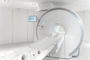
What Is an MRI?
Magnetic resonance imaging (MRI) is a medical imaging technique that uses a powerful magnetic field and radio waves to create detailed images of the body’s internal structures. Unlike X-rays and CT scans, which use ionising radiation, an MRI is noninvasive and does not expose patients to harmful radiation.
During an MRI, you will lie on a table that slides into a tunnel-like machine. The machine generates a magnetic field and radio waves, which are picked up by a receiver and used to create detailed images of your shoulder. The entire procedure usually takes around 20 to 30 minutes.
What is an MRI arthrogram and when is it used?
The MRI is a highly accurate diagnostic tool that can provide detailed images of the shoulder joint, including the bones, cartilage, tendons and ligaments. It provides a good level of detail, but occasionally you may be asked to have an arthrogram as well. This is particularly good for imaging a shoulder with instability symptoms.
An arthrogram is an injection of dye contrast, placed directly into the joint followed immediately by the MRI. The benefit of the dye is to assess the volume of the shoulder joint capsule and subtle splits or tears to the rim of the socket that might otherwise be missed on a plain MRI.
The injection is usually performed by a radiologist (doctor) and using an ultrasound or X-ray machine so that the needle is placed accurately in the joint. It is done as an outpatient procedure.
How to prepare for an MRI scan
If you need an MRI scan for shoulder pain, it is important to follow any preparation instructions provided by the clinic. This may include removing any metal objects from your body, and wearing loose, comfortable clothing.
Arrive in plenty of time for your appointment as you may be required to fill in a form and get changed into a hospital gown.
Some people find the MRI tube claustrophobic and I recommend wearing an eye-mask so that you can not see the machine when you are inside.
The machine will make some very loud noises during the test, this is normal and nothing to be concerned about. Most imaging centres offer you a headset so that you can choose to listen to music or a radio station whilst you are in the scanner.
Remember to breathe slowly and deeply to help you relax. Think about something that makes you feel happy and remember that it will only last for 20-30 minutes and you will soon be out. You will be given a remote button to press so that if at any stage you feel that you need to get out, the radiographers will immediately remove you safely from the scanner.
They have a window that looks directly into the scanning room so you will be watched at all times and are completely safe.
Treatment Planning with MRI
One of the main benefits of an MRI scan for shoulder pain is for developing an effective and customised treatment plan based on the specific condition or injury. For example, if you have a rotator cuff tear, an MRI scan helps determine the size and location of the tear, as well as the extent of muscle and tissue damage. With this information, I can develop a customised treatment plan that may include physical therapy, medication, injection therapy or surgery.
Conclusion
If you are experiencing persistent shoulder pain, an MRI scan may be necessary to obtain an accurate diagnosis and develop an effective treatment plan. MRI is a safe and non-invasive procedure that can provide detailed images of the soft tissues, bones and blood vessels inside the shoulder joint. It is particularly useful for detecting soft tissue injuries and abnormalities, such as rotator cuff tears and labral tears. If you are unsure whether an MRI is necessary for your shoulder pain, come and have a consultation with me and we can discuss the different options available to you.
It is worth noting that an MRI scan may not be necessary for every case of shoulder pain. In some cases, rest, physical therapy and other non-invasive treatments may be sufficient to alleviate pain and improve shoulder function. However, if your symptoms persist despite treatment, or if they are severe, an MRI scan may be recommended.
Overall, an MRI scan is an important tool in the diagnosis and treatment of shoulder pain. By providing detailed images of the shoulder joint and surrounding tissues, MRI can help doctors develop an accurate diagnosis and treatment plan, ultimately leading to improved outcomes for patients.
Introducing Your Shoulder
Your Shoulder is the online practice of Susan Alexander, a shoulder orthopaedic surgeon with over 20 years of experience in treating shoulder injuries. You can find more information about Susan at Your Shoulder’s About Me page.
If you’re experiencing shoulder pain and are wondering if and when you may need an MRI scan, email or call Your Shoulder on +44 (0) 203 195 2459 to book an appointment.
For more information on shoulder problems, visit the How I Can Help Page at Your Shoulder.
recommend an MRI scan to obtain a more detailed image of your shoulder joint and the surrounding tissues. MRI can help your doctor determine the extent of the injury or condition and develop an effective treatment plan.
Client testimonial:
Susan is amazing. Not only did she transform my shoulder with the surgery, but her frank and helpful approach meant I was kept really informed throughout. She also places a real focus on rehabilitation, recommending an excellent physio to me but also recommending particular exercises (even demonstrating them herself to ensure I understood them correctly).
Ms Susan Alexander is a Consultant Orthopaedic Surgeon and President of the Independent Doctors Federation.
She specialises in all shoulder conditions, including frozen shoulder, rotator cuff tears, dislocations and arthritis. Based in London, she practises at Fortius Clinic and King Edward VII’s Hospital. Ms Alexander is highly experienced in minimally invasive (keyhole) shoulder surgery and is known for her meticulous, patient-centred and holistic approach.
She focuses on accurate diagnosis, bespoke treatment planning, and ongoing support throughout recovery. Learn more about Ms Susan Alexander here.


